

In this article we will explore what exactly electroencephalography is, what it is used for, and who uses it. Common questions about electroencephalography will also be answered, including a mini deep-dive into the technology used.
What is Electroencephalography?
Electroencephalography (EEG) is a non-invasive technique to record electrical activity in the brain. Electrodes are placed on the scalp to measure brain waves. Electroencephalography helps diagnose neurological disorders and evaluate brain function, and is also used in research to study brain activity.
How do you pronounce Electroencephalography?
Daunting as it might first appear, it is easily broken down into short phonetic sounds.
Try saying:
ELEC – TRO – EN – SEFFA – LOG – RAFFY
You can hear it being spoken over on youtube.
Who Discovered Electroencephalography?
The discovery of EEG can be attributed to Hans Berger, a German psychiatrist and neurologist.
Berger became interested in the study of the electrical activity of the brain, which was a relatively new field at the time. He conducted experiments using electrodes placed on the scalp of human volunteers and made his first EEG recording in 1924 on a 17 year-old boy undergoing neurosurgery. Berger observed that the electrical activity of the brain varied according to the state of consciousness of the subject, and he concluded that these variations could be used to diagnose neurological disorders.
Berger published his findings in 1929 in a paper entitled “Über das Elektrenkephalogramm des Menschen” (On the Electroencephalogram of Man)1. The paper described Berger’s experiments and provided evidence of the existence of alpha waves, which are the dominant electrical signals produced by the brain when a person is awake and relaxed.
Berger’s discovery of EEG revolutionized the field of neuroscience and has led to numerous advances in the diagnosis and treatment of neurological disorders. Today, EEG is widely used in clinical settings to diagnose and monitor conditions such as epilepsy, sleep disorders, and brain injuries.
Hans Berger was a ground-breaking researcher who has had a profound impact on the field of neuroscience and has contributed significantly to our understanding of the human brain.
How is Electroencephalography Recorded?
Electroencephalography is commonly used to help diagnose and monitor epilepsy. Clinicians will engage patients with various cognitive and behavioural tasks whilst monitoring their brain activity.
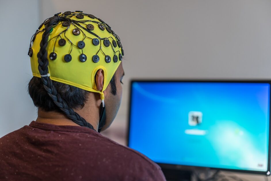
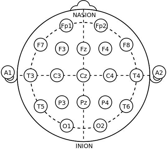
During an EEG recording, electrodes are placed on the scalp using a conductive gel. The electrodes detect electrical activity in the brain and transmit the signal to an amplifier. The amplified signal is then sent to a computer for analysis. The placement of the electrodes is important for obtaining accurate recordings, and the International 10-20 System is commonly used to ensure consistency across studies and institutions. For more information on the 10-20 system, read this article.
Electroencephalography recordings are typically performed in a quiet and dimly lit room to minimise environmental interference. Patients are usually asked to sit or lie down comfortably and remain still during the recording, as movement can create artifacts in the signal. EEG recordings can last from a few minutes to several hours, depending on the purpose of the study.
Are There Different Types of EEG?
There are various types of EEG recordings, including routine EEG, ambulatory EEG, and video EEG monitoring.
Routine EEG is a brief recording (typically 20-30 minutes) performed in a clinical setting to diagnose and monitor epilepsy.
Ambulatory EEG involves wearing a portable EEG recorder for several days to capture seizure activity outside of the clinical setting.
Video EEG monitoring combines EEG recording with video surveillance to provide a more comprehensive assessment of seizure activity.
In research, diagnosis of neurodegenerative diseases is combining EEG with with functional magnetic resonance imaging (fMRI). Traditionally fMRI was used instead of, and as a superior alternative to EEG due to its enhanced spatial resolution. EEG however can offer much greater temporal resolution, and therefore research groups are now looking at combining the methods for more advanced diagnosis2.
Conclusion
In conclusion, EEG recording is a non-invasive technique that involves placing electrodes on the scalp to detect electrical activity in the brain. EEG recordings are important for diagnosing and monitoring epilepsy, as well as for studying brain activity in various cognitive and behavioural tasks. The placement of the electrodes and the recording conditions are crucial for obtaining accurate recordings.
Continued research into EEG is helping to develop more sophisticated tools not only for diagnosis in the clinical setting, but also to help Neurologists and Psychiatrists develop better understanding about how the human brain works.
For more information on other types of biosignals, you may be interested in reading about electrocardiography (ECG) or photoplethysmography (PPG). We also have more biomedical engineering articles you may be interested in here.
References
- H. Berger, ‘Über das Elektrenkephalogramm des Menschen’, Archiv f. Psychiatrie, vol. 87, no. 1, pp. 527–570, Dec. 1929, doi: 10.1007/BF01797193.
- G. Mele, C. Cavaliere, V. Alfano, M. Orsini, M. Salvatore, and M. Aiello, ‘Simultaneous EEG-fMRI for Functional Neurological Assessment’, Frontiers in Neurology, vol. 10, 2019, Accessed: Apr. 13, 2023. [Online]. Available: https://www.frontiersin.org/articles/10.3389/fneur.2019.00848